
Plantar Fasciitis Myofascial Connections and Yoga
The therapeutic benefits of Hatha yoga arise from whole body energetic balancing combined with distinct biomechanical adjustments. We gave an example of this in our last blog post, where we looked at the disorder known as adult acquired flatfoot deformity, its biomechanical basis and how to utilize yoga to maintain healthy foot arches. In this post we focus on the plantar fascia of the foot and examine the most common cause of heel pain plantar fasciitisto see what happens when things go wrong. Finally, we consider how yoga can be used to bring things back into balance and even to prevent this condition. First, let’s look at fascia in general.
A fascia is a fibrous structure that is formed from sheets of connective tissue. The deep fascia covers and invests muscles, tendons, ligaments and blood vessels throughout the body. An important example of a deep fascia is the thoracolumbar fascia. All yoga practitioners should be familiar with this structure and its myofascial connections, as it forms a critical support system for the lumbar spine and sacroiliac joint. Other types of fascia include the superficial fascia of the subcutaneous tissue under the skin, and the visceral andparietal fascia, which surround the organs such as the heart and lungs. Figure 1 illustrates the deep fascial elements of skeletal muscles.
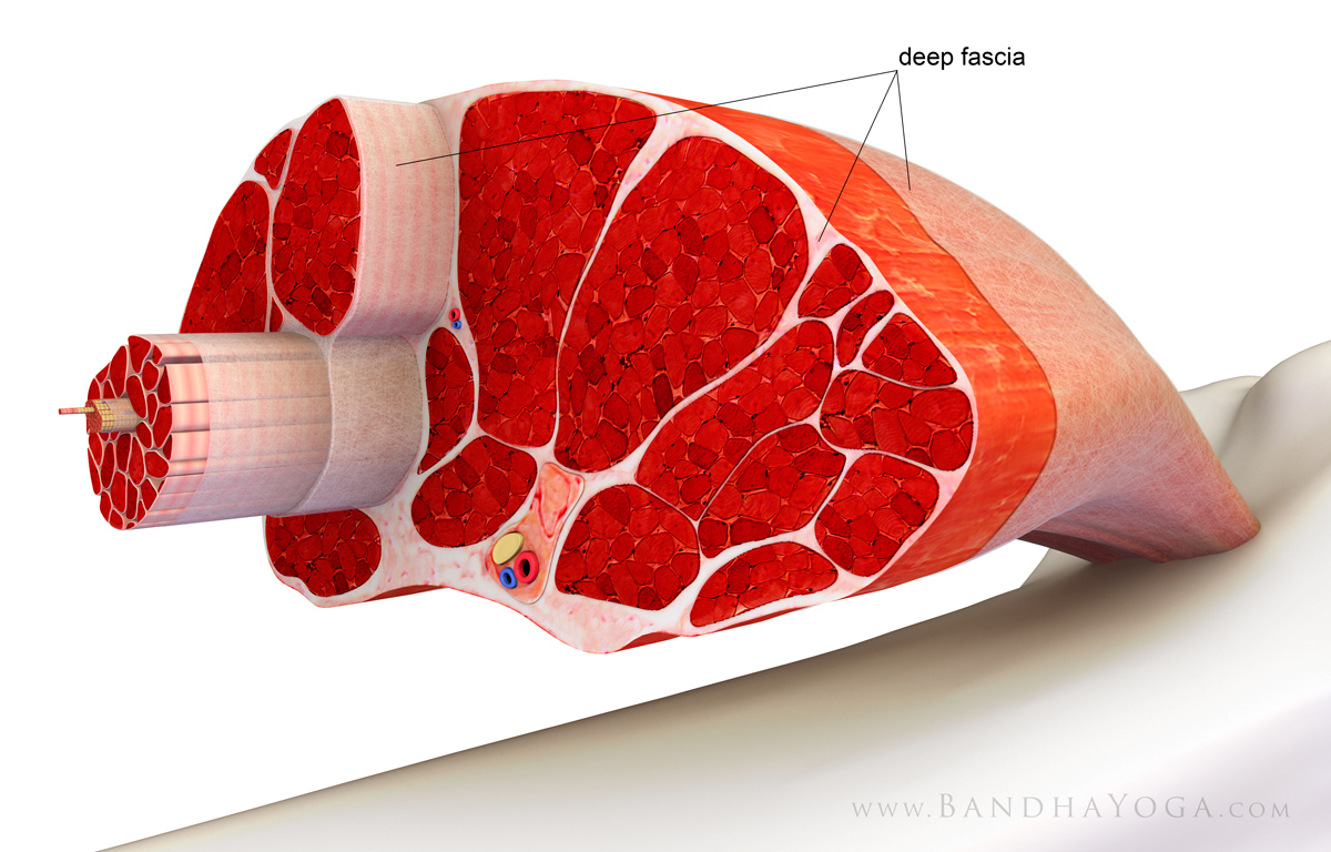
Figure 1 The deep fascia covering and investing skeletal muscle.
The plantar fascia or plantar aponeurosis you can use either term originates from the medial tubercle of the calcaneus heel bone and continues forward to attach to the proximal phalanx of each of the toes via the plantar plates. Extending dorsiflexing the toes tightens the plantar fascia, thus elevating the foot arch. During this process, the metatarsal heads act as pulleys to form a windlass to tighten the plantar aponeurosis. The plantar fascia has elastic qualities in that its fibers are somewhat wavy in the relaxed position. These fibers straighten in response to forces applied like the heel-off phase of gait. Thus, the plantar fascia can store energy like a spring. Figure 2 illustrates this concept.
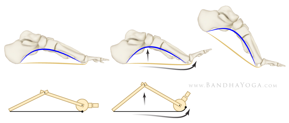
Figure 2: The windlass mechanism of the plantar aponeurosis fascia.
The plantar aponeurosis also forms a myofascial connection with the muscles of the calf gastrocnemius and soleus via the Achilles tendon and, by extension, the hamstrings and potentially other muscles of the posterior kinetic chain. Forces that stretch the plantar fascia are distributed along these muscles. Conversely, tightness in these muscles can adversely affect the function of the plantar fascia and thus the arch of the foot. Figure 3 illustrates these myofascial connections in Downward Facing Dog pose.
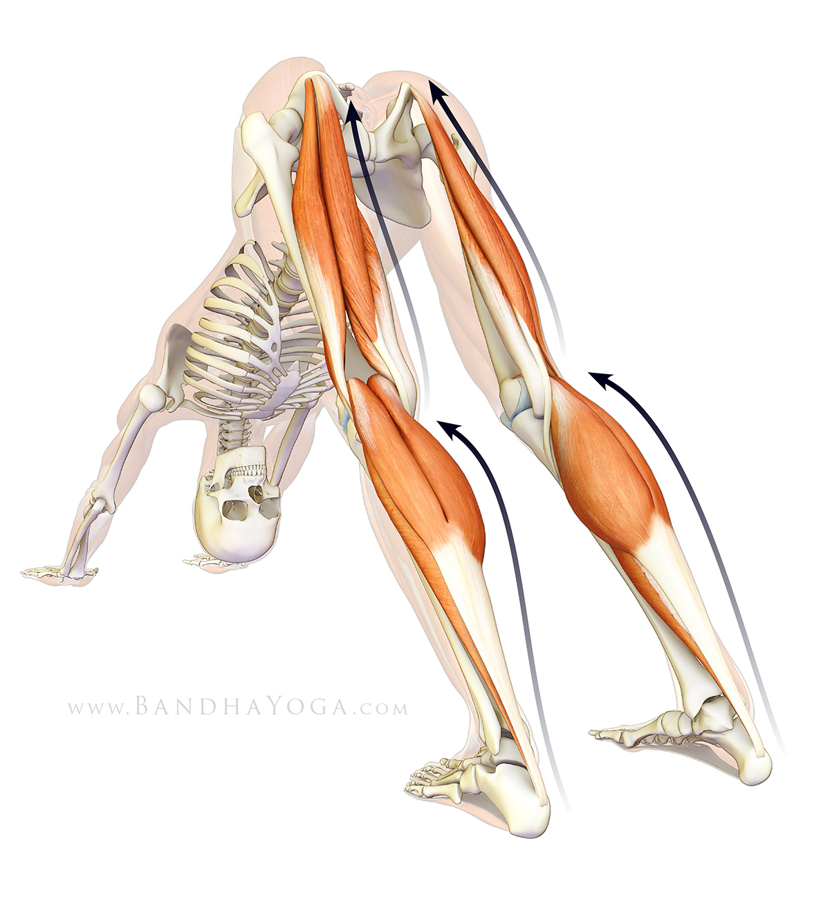
Figure 3 The myofascial connections to the plantar fascia in Downward Dog pose.
Plantar fasciitis is an overuse injury related to repetitive overstretching of the plantar aponeurosis. In this condition the forces of gait are concentrated where the plantar fascia attaches to the calcaneus, instead of being distributed over the muscles at the back of the legs. This results in microtrauma to the plantar aponeurosis near its origin, causing inflammation and heel pain. Risk factors for developing plantar fasciitis include tight calf muscles and hamstrings, endurance-type weight bearing activity such as running and a high body mass index. Figure 4 illustrates plantar fasciitis.
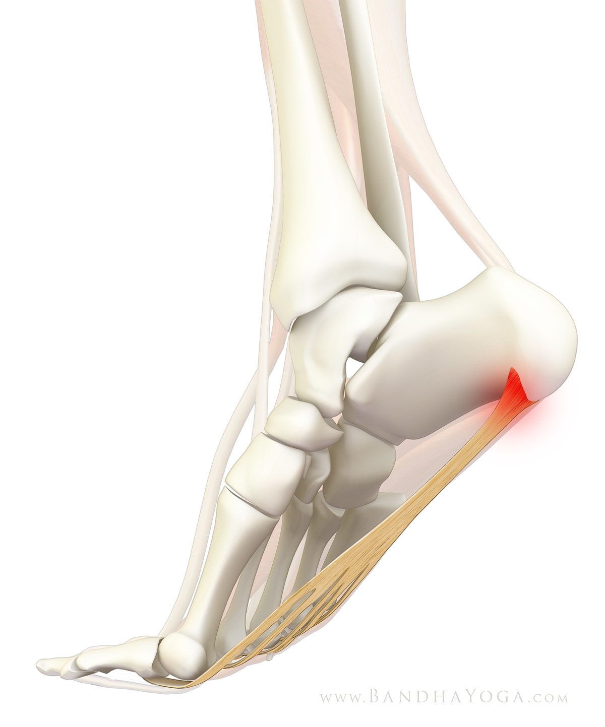
Figure 4: Plantar fasciitis note the inflammation at the origin of the plantar aponeurosis.
Note that there are other conditions that can cause heel pain. An example of such a condition is a stress fracture of the calcaneus, which is also seen in runners. This problem is treated differently from plantar fasciitis. Accordingly, if you have heel pain be sure to consult a health care practitioner who is appropriately trained and qualified to diagnose and manage such conditions. Use your knowledge of pathological conditions to deepen your understanding of the body and to work with yoga as an adjunct in prevention and treatment.
Since the main means of managing this condition is stretching of the plantar fascia, heel cords gastrocnemius/soleus complex and hamstrings, yoga offers an ancient preventative solution. For example, Downward Dog pose stretches both the hamstrings and heel cords.
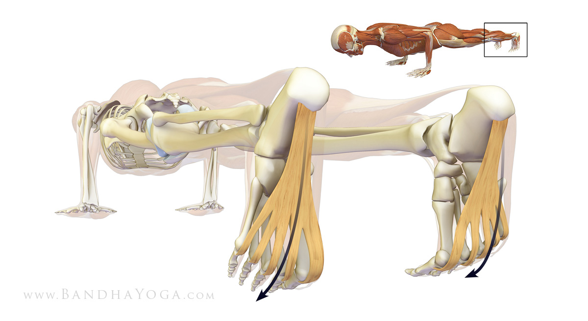
Figure 5: Stretching the plantar aponeurosis fascia in Chaturanga dandasana.
Chaturanga dandasana figure 5stretches the plantar fascia itself. Use this image to aid in visualization of this process while in the pose. One of our previous posts gives some tips on how to ease into Chaturanga and another describes a key muscular co-contraction in this pose.
Uttanasana illustrates a stretch of the posterior kinetic chain, linking to the feet figure 6. Click here for a tip on integrating the hip abductors to access sacral nutation to refine Uttanasana. Thus, we can see that the Sun Salutations Surya Namaskar offer an ancient self-contained method for working with the plantar fascia and its myofascial connections to maintain a healthy foot arch. For many more tips and cues like this, check out the Yoga Mat Companion book series and The Key Muscles and Key Poses of Yoga.
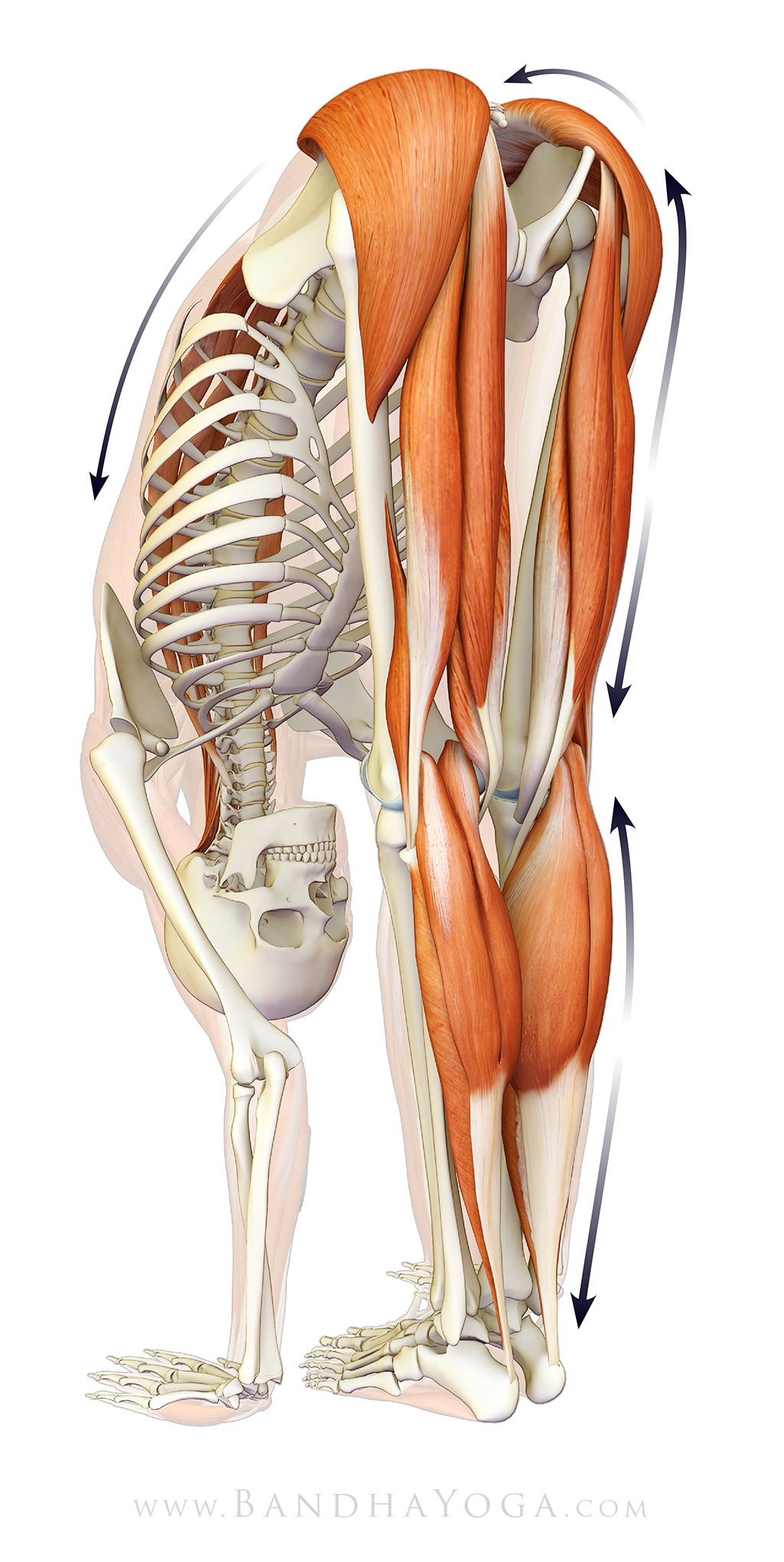
Figure 6: The posterior kinetic chain and its connection to the feet in Uttanasana.
We conclude with a step-wise technique on using biomechanics and physiology to lengthen the heel cords in Janu Sirsana (seated forward bend):
Step one: Bend the knee about 15 degrees to release the gastrocnemius muscle at its origin on the posterior femur.
Step two: Use the hands to gently draw the ankle into dorsiflexion and stabilize it in this position by engaging the biceps to flex the elbows. The cue I use for this is to "draw the top of the foot towards the front of the shin (dorsiflexion)."
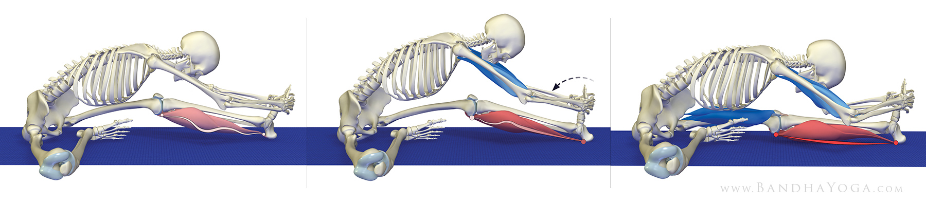
Figure 7: Steps to release and then lengthen the calf muscles in Janu sirsasana.
Step three: Hold the foot in place and gradually engage the quadriceps to straighten the knee. Ease into this position. Maintaining the ankle in some dorsiflexion with the arms and extending the knee distributes the stretch throughout the calf muscles (the gastrocnemius and soleus) as illustrated here.
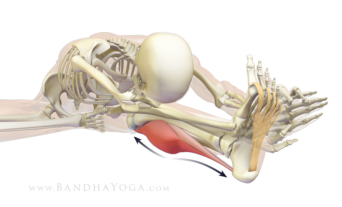
Figure 8 The myofascial connection between the plantar fascia, heel cord and calf muscles.
You can add a facilitated stretch to the calf by gently pressing the ball of the foot into the hands for 8-10 seconds and then taking up the slack by further dorsiflexing the ankle. This activates the Golgi tendon organ at the muscle tendon junction, resulting in relaxation of the contractile elements. We describe a similar technique to lengthen the hamstrings, as well as the physiological basis for facilitated stretching in our blog post how to lengthen the hamstrings in Janu sirsasana.
The therapeutic benefits of Hatha yoga arise from whole body energetic balancing combined with distinct biomechanical adjustments. We gave an example of this in our last blog post, where we looked at the disorder known as adult acquired flatfoot deformity, its biomechanical basis and how to utilize yoga to maintain healthy foot arches. In this post we focus on the plantar fascia of the foot and examine the most common cause of heel pain plantar fasciitisto see what happens when things go wrong. Finally, we consider how yoga can be used to bring things back into balance and even to prevent this condition. First, let’s look at fascia in general.
A fascia is a fibrous structure that is formed from sheets of connective tissue. The deep fascia covers and invests muscles, tendons, ligaments and blood vessels throughout the body. An important example of a deep fascia is the thoracolumbar fascia. All yoga practitioners should be familiar with this structure and its myofascial connections, as it forms a critical support system for the lumbar spine and sacroiliac joint. Other types of fascia include the superficial fascia of the subcutaneous tissue under the skin, and the visceral andparietal fascia, which surround the organs such as the heart and lungs. Figure 1 illustrates the deep fascial elements of skeletal muscles.
Figure 1 The deep fascia covering and investing skeletal muscle.
The plantar fascia or plantar aponeurosis you can use either term originates from the medial tubercle of the calcaneus heel bone and continues forward to attach to the proximal phalanx of each of the toes via the plantar plates. Extending dorsiflexing the toes tightens the plantar fascia, thus elevating the foot arch. During this process, the metatarsal heads act as pulleys to form a windlass to tighten the plantar aponeurosis. The plantar fascia has elastic qualities in that its fibers are somewhat wavy in the relaxed position. These fibers straighten in response to forces applied like the heel-off phase of gait. Thus, the plantar fascia can store energy like a spring. Figure 2 illustrates this concept.
Figure 2: The windlass mechanism of the plantar aponeurosis fascia.
The plantar aponeurosis also forms a myofascial connection with the muscles of the calf gastrocnemius and soleus via the Achilles tendon and, by extension, the hamstrings and potentially other muscles of the posterior kinetic chain. Forces that stretch the plantar fascia are distributed along these muscles. Conversely, tightness in these muscles can adversely affect the function of the plantar fascia and thus the arch of the foot. Figure 3 illustrates these myofascial connections in Downward Facing Dog pose.
Figure 3 The myofascial connections to the plantar fascia in Downward Dog pose.
Plantar fasciitis is an overuse injury related to repetitive overstretching of the plantar aponeurosis. In this condition the forces of gait are concentrated where the plantar fascia attaches to the calcaneus, instead of being distributed over the muscles at the back of the legs. This results in microtrauma to the plantar aponeurosis near its origin, causing inflammation and heel pain. Risk factors for developing plantar fasciitis include tight calf muscles and hamstrings, endurance-type weight bearing activity such as running and a high body mass index. Figure 4 illustrates plantar fasciitis.
Figure 4: Plantar fasciitis note the inflammation at the origin of the plantar aponeurosis.
Note that there are other conditions that can cause heel pain. An example of such a condition is a stress fracture of the calcaneus, which is also seen in runners. This problem is treated differently from plantar fasciitis. Accordingly, if you have heel pain be sure to consult a health care practitioner who is appropriately trained and qualified to diagnose and manage such conditions. Use your knowledge of pathological conditions to deepen your understanding of the body and to work with yoga as an adjunct in prevention and treatment.
Since the main means of managing this condition is stretching of the plantar fascia, heel cords gastrocnemius/soleus complex and hamstrings, yoga offers an ancient preventative solution. For example, Downward Dog pose stretches both the hamstrings and heel cords.
Figure 5: Stretching the plantar aponeurosis fascia in Chaturanga dandasana.
Chaturanga dandasana figure 5stretches the plantar fascia itself. Use this image to aid in visualization of this process while in the pose. One of our previous posts gives some tips on how to ease into Chaturanga and another describes a key muscular co-contraction in this pose.
Uttanasana illustrates a stretch of the posterior kinetic chain, linking to the feet figure 6. Click here for a tip on integrating the hip abductors to access sacral nutation to refine Uttanasana. Thus, we can see that the Sun Salutations Surya Namaskar offer an ancient self-contained method for working with the plantar fascia and its myofascial connections to maintain a healthy foot arch. For many more tips and cues like this, check out the Yoga Mat Companion book series and The Key Muscles and Key Poses of Yoga.
Figure 6: The posterior kinetic chain and its connection to the feet in Uttanasana.
We conclude with a step-wise technique on using biomechanics and physiology to lengthen the heel cords in Janu Sirsana (seated forward bend):
Step one: Bend the knee about 15 degrees to release the gastrocnemius muscle at its origin on the posterior femur.
Step two: Use the hands to gently draw the ankle into dorsiflexion and stabilize it in this position by engaging the biceps to flex the elbows. The cue I use for this is to "draw the top of the foot towards the front of the shin (dorsiflexion)."
Figure 7: Steps to release and then lengthen the calf muscles in Janu sirsasana.
Step three: Hold the foot in place and gradually engage the quadriceps to straighten the knee. Ease into this position. Maintaining the ankle in some dorsiflexion with the arms and extending the knee distributes the stretch throughout the calf muscles (the gastrocnemius and soleus) as illustrated here.
Figure 8 The myofascial connection between the plantar fascia, heel cord and calf muscles.
You can add a facilitated stretch to the calf by gently pressing the ball of the foot into the hands for 8-10 seconds and then taking up the slack by further dorsiflexing the ankle. This activates the Golgi tendon organ at the muscle tendon junction, resulting in relaxation of the contractile elements. We describe a similar technique to lengthen the hamstrings, as well as the physiological basis for facilitated stretching in our blog post how to lengthen the hamstrings in Janu sirsasana.
From the group at Bandha Yoga Bless you guys great work Bud
No comments:
Post a Comment